
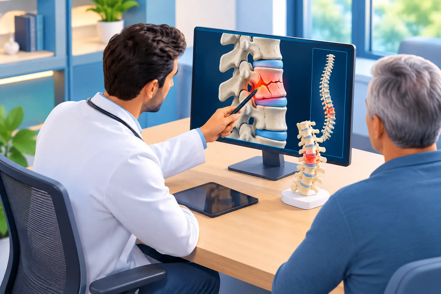
A vertebral fracture can turn an ordinary movement – reaching for a shirt, standing from a chair, taking a short walk – into a sharp reminder that the spine needs attention. To manage vertebral fractures well, the first priority is determining what caused the break, whether the spine is stable, and how pain is affecting safe movement. The right plan is rarely one-size-fits-all. It should protect the injured vertebra while helping the patient remain as mobile and independent as possible.
For many adults, especially those with osteoporosis, a vertebral compression fracture may occur after a minor fall, an awkward movement, or no clearly memorable injury at all. Others result from higher-impact trauma. In either case, prompt evaluation matters because untreated fractures can lead to prolonged pain, reduced activity, posture changes, and additional stress on the surrounding spine.
When Vertebral Fracture Symptoms Need Urgent Care
Back pain after a fall, collision, or sudden strain deserves medical attention when it is severe, persistent, or makes standing and walking difficult. A new fracture can cause focal pain that worsens with movement, coughing, or changing positions. Some patients also notice a loss of height or a more rounded upper-back posture over time.
Certain symptoms require emergency assessment rather than waiting for an office visit. These include new weakness or numbness in the legs, loss of bowel or bladder control, numbness in the groin area, difficulty walking, fever with severe back pain, or pain following a significant accident. These signs can indicate nerve involvement, infection, or an unstable injury that needs immediate care.
Not every painful back episode is a fracture. Arthritis, muscle injury, disc conditions, spinal stenosis, and cancer-related spine changes can create overlapping symptoms. That is why a careful diagnosis is essential before choosing treatment.
How Specialists Manage Vertebral Fractures Safely
A focused evaluation begins with the patient’s symptoms, injury history, medical conditions, medications, and bone-health risk factors. Imaging may include X-rays, CT scans, or MRI. Each answers a different question. X-rays can show vertebral height loss and alignment; CT provides detailed bone anatomy; MRI can help determine whether a fracture is new, identify swelling in the bone, and evaluate nerves and soft tissues.
The results help distinguish a stable compression fracture from an injury that may require surgical consultation or more urgent stabilization. They also help identify the source of pain. A vertebra that appears compressed on an X-ray may represent an older healed fracture rather than the cause of a patient’s current symptoms.
At Global Pain Solutions, treatment planning is built around that level of precision. The goal is not simply to quiet pain temporarily. It is to identify the pain generator, protect function, and select the least invasive treatment that fits the patient’s condition and recovery goals.
Initial treatment for stable fractures
Many stable vertebral compression fractures improve without major surgery. Early care may involve a limited period of activity modification, with guidance on avoiding heavy lifting, repeated bending, twisting, and high-impact exercise while the bone heals. Complete bed rest is usually not the answer. Extended inactivity can weaken muscles, increase stiffness, worsen bone loss, and raise the risk of blood clots or falls.
A brace may be appropriate for some patients. It can limit painful motion and provide support during the early healing period, particularly when getting up, walking, or completing daily tasks. Bracing has trade-offs, however. Wearing one too long can contribute to trunk muscle weakness, skin irritation, and discomfort. The duration and type of brace should be individualized rather than treated as a default solution.
Medication may also have a role, but it should be used thoughtfully. Acetaminophen or anti-inflammatory medication may help certain patients, depending on kidney function, stomach health, cardiovascular risk, and other medications. Short-term medication for severe acute pain may sometimes be necessary, but long-term opioid reliance does not address fracture healing, bone strength, or the mechanics driving ongoing discomfort.
As pain begins to settle, carefully directed physical therapy can help restore safe mobility, posture, balance, and spinal support. The timing matters. Exercises that are helpful later in recovery may be too provocative during the acute phase, so a progressive plan is safer than pushing through severe pain.
When Kyphoplasty May Be Considered
For some patients, pain remains intense despite appropriate conservative care and substantially limits walking, sleep, self-care, or return to work. When imaging confirms a painful, recent compression fracture, kyphoplasty may be considered.
Kyphoplasty is a minimally invasive procedure designed to stabilize a compressed vertebra. Through small incisions and image guidance, a physician creates space within the fractured bone and places bone cement to provide internal support. For appropriately selected patients, the procedure may reduce fracture-related pain and allow earlier return to movement.
It is not the right treatment for every vertebral fracture. The decision depends on the age and pattern of the fracture, the patient’s neurological status, overall health, degree of vertebral collapse, response to conservative treatment, and whether the painful level is clearly identified. There are also procedural risks, including bleeding, infection, cement leakage, and complications related to sedation, although careful technique and patient selection are intended to minimize them.
A meaningful conversation should address both the potential benefits and the alternatives. Some patients improve steadily with bracing, medication, and rehabilitation. Others need a more targeted intervention because pain has stalled recovery. Personalized guidance is especially valuable when the goal is to regain function without unnecessary surgery.
Treat the Bone Health Problem Behind the Break
A vertebral compression fracture is often more than an isolated spine injury. In adults over 50, particularly women after menopause and men with known risk factors, it can be a warning sign of osteoporosis or reduced bone density. One fracture increases the likelihood of another, which makes prevention part of fracture care.
Bone health evaluation may include a bone-density test, review of calcium and vitamin D intake, laboratory testing when indicated, and assessment for medical causes of bone loss. Conditions involving thyroid function, kidney disease, digestive disorders, inflammatory illness, low hormone levels, and certain medications can affect bone strength. Long-term corticosteroid use is a well-known risk factor.
Treatment may involve nutrition changes, weight-bearing activity when medically cleared, fall-prevention strategies, and prescription osteoporosis medication through the appropriate clinician. The best approach depends on fracture history, bone-density results, kidney function, and an individual’s broader medical picture. Simply taking a supplement without evaluating the cause of bone loss may leave a major risk unaddressed.
Movement, Home Safety, and Recovery Expectations
Recovery varies. Some uncomplicated compression fractures improve significantly over several weeks, while others take months to settle fully. Patients with multiple fractures, severe osteoporosis, preexisting spinal degeneration, or chronic pain may need a longer and more structured recovery plan.
During recovery, small practical changes can reduce the chance of another injury. Supportive footwear, clear walking paths, adequate home lighting, and attention to vision or balance problems all matter. If dizziness or medication side effects contribute to falls, those issues should be reviewed with a medical professional.
Patients should also avoid interpreting pain relief as an immediate return to unrestricted activity. Healing bone still needs protection. A clinician can help determine when it is reasonable to resume lifting, golf, fitness training, travel, or work duties that involve repetitive bending or long periods on the feet.
Persistent pain after a fracture does not automatically mean the bone failed to heal. It may reflect muscle guarding, altered movement patterns, facet-joint irritation, nerve compression, or another spinal condition that became more noticeable during recovery. Reassessment can identify whether a targeted interventional treatment, rehabilitation adjustment, or another specialist evaluation is appropriate.
A vertebral fracture can be disruptive, but it does not have to define a patient’s future mobility. Timely diagnosis, thoughtful pain care, and attention to bone health create the strongest path back to safer movement and greater confidence in daily life.